Abstract
Ventricular fibrillation (VF) is a life-threatening electromechanical dysfunction of the heart associated with complex spatiotemporal dynamics of electrical excitation and mechanical contraction of the heart muscle. It has been hypothesized that VF is driven by three-dimensional rotating electrical scroll waves, which can be characterized by filamentlike electrical phase singularities or vortex filaments, but visualizing their dynamics has been a long-standing challenge. Recently, it was shown that rotating excitation waves during VF are associated with rotating waves of mechanical deformation. Three-dimensional mechanical scroll waves and mechanical filaments describing their rotational core regions were observed in the ventricles by using high-resolution ultrasound. The findings suggest that the spatiotemporal organization of cardiac fibrillation may be assessed from waves of mechanical deformation. However, the complex relationship between excitation and mechanical waves during VF is currently not understood. Here, we study the fundamental nature of mechanical phase singularities, their spatiotemporal organization, and their relation with electrical phase singularities. We demonstrate the existence of two fundamental types of mechanical phase singularities: “paired singularities,” which are colocalized with electrical phase singularities, and “unpaired singularities,” which can form independently. We show that the unpaired singularities emerge due to the anisotropy of the active force field, generated by fiber anisotropy in cardiac tissue, and the nonlocality of elastic interactions, which jointly induce strong spatiotemporal inhomogeneities in the strain fields. The inhomogeneities lead to the breakup of deformation waves and create mechanical phase singularities, even in the absence of electrical singularities, which are typically associated with excitation wave break. We exploit these insights to develop an approach to discriminate paired and unpaired mechanical phase singularities, which could potentially be used to locate electrical rotor cores from a mechanical measurement. Our findings provide a fundamental understanding of the complex spatiotemporal organization of electromechanical waves in the heart and a theoretical basis for the analysis of high-resolution ultrasound data for the three-dimensional mapping of heart rhythm disorders.
- Received 14 July 2021
- Revised 22 February 2022
- Accepted 18 April 2022
DOI:https://doi.org/10.1103/PhysRevX.12.021052
Published by the American Physical Society under the terms of the Creative Commons Attribution 4.0 International license. Further distribution of this work must maintain attribution to the author(s) and the published article’s title, journal citation, and DOI.
Published by the American Physical Society
Physics Subject Headings (PhySH)
Popular Summary
A normal heartbeat is orchestrated by the propagation of an electrical wave that triggers a synchronized mechanical contraction of the heart muscle. During life-threatening cardiac arrhythmias, in which something alters this steady rhythm, the electrical waves can become disorganized and stop the heart from pumping. Here, we demonstrate computationally that, in this setting, waves of mechanical contraction do not simply follow excitation waves but rather exhibit a higher degree of spatiotemporal disorganization.
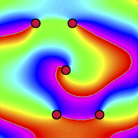
Our work relies on phase mapping, a powerful tool for visualizing the progression of excitations and tissue deformation across the heart muscle. During a normal heartbeat, waves propagate across the heart in straight lines; at each point of the phase map, that propagation resembles a hand going around a clock, and the phase is continuous in space. During arrhythmias, however, excitation waves rapidly rotate around a singular point where the phase cannot be defined—as at the center of a clock.
Our results demonstrate that, in addition to these singular points associated with rotating excitation waves, phase maps of mechanical waves contain other singular points as well. This is because phase maps of electrical excitation and tissue deformation do not necessarily coincide. Electrical excitation signals a cell to contract, but the resulting force can deform tissue far from the point of excitation.
These results provide an improved basis for using high-resolution ultrasound images of heart-contraction waves to infer excitation-wave patterns underlying clinically important cardiac arrhythmias.